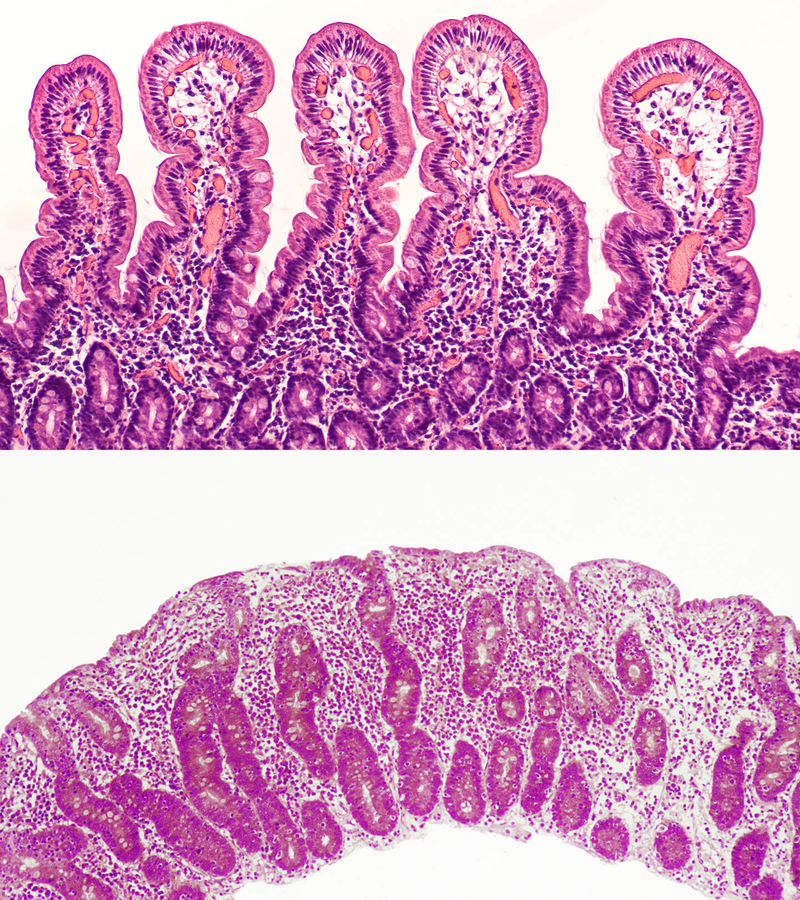
Evidence #4: Small Intestine Micrographs
How does food move from the small intestine to the rest of the body, and what is different about M'Kenna's intestine?
Courtesy David Litman/Shutterstock.com, Biophoto Associates/Science Photo Library
When M'Kenna visited her doctor for the endoscopy exam, the doctor decided to take a sample of tissue to examine under a microscope. The sample of tissue was very small. The doctor took the sample to the lab and looked at it through a microscope.
The doctor then made a photograph of the sample to show M'Kenna and her family. A photograph taken by a microscope is called a micrograph. In this activity, you will examine two micrographs and make observations of the tissue in both pictures. You may even be able to identify cells in the tissue you see. You will decide which tissue you believe is M'Kenna's and why you think the tissue is not doing a good job absorbing nutrients.
Collaboration Icon
Work with your team to make observations of micrographs.
With your team
- Make observations of the two micrographs. Use the Observations of Intestinal Tissue handout to record your observations.
- Make a claim about which tissue belongs to M'Kenna and use evidence from your observations to support your claim.
- Be prepared to share what you observed and your claim when prompted by your teacher.
On your own
- Attach Observations of Intestinal Tissue to your science notebook. Title the page, "Evidence #4: Small Intestine Micrographs."
